Mesothelioma Histology Pathology Outlines / Pathology Outlines Mesothelial : Distinct nucleoli may be present.. Ttf1, pax8, etc.) use more markers if results inconclusive best positive mesothelioma markers. 02.07.2021 · the most common histologic patterns of epithelioid mesothelioma are tubulopapillary, adenomatoid, solid well differentiated, solid poorly differentiated and acinar ( … Mesothelioma pathology provides a full picture of the cancer, contributing to a more accurate diagnosis and an informed … Mesothelial cells form conspicuous layer of regularly spaced, bland cuboidal cells along pleural surface; Papillae with myxoid cores, each lined by a …
Papillae with myxoid cores, each lined by a … Distinct nucleoli may be present. Mesothelial cells form conspicuous layer of regularly spaced, bland cuboidal cells along pleural surface; Mesothelioma pathology provides a full picture of the cancer, contributing to a more accurate diagnosis and an informed … Mesothelioma histology, or mesothelioma histopathology, is the study of tissue for the presence of mesothelioma.
1 from Ttf1, pax8, etc.) use more markers if results inconclusive best positive mesothelioma markers. 04.05.2021 · international mesothelioma interest group recommends the initial diagnostic immunohistochemical panel should include at least 2 mesothelial markers and 2 general epithelial markers for other tumors in the differential diagnosis, on the basis of morphology, specific markers can be added (e.g. Mesothelioma pathology provides a full picture of the cancer, contributing to a more accurate diagnosis and an informed … This process is part of mesothelioma … 03.03.2021 · malignant mesothelioma in situ (histopathology 2018;72:1033): Papillae with myxoid cores, each lined by a … 23.05.2019 · well differentiated papillary mesothelioma. Most commonly in the peritoneum, rarely pleura and other sites histology:
04.05.2021 · international mesothelioma interest group recommends the initial diagnostic immunohistochemical panel should include at least 2 mesothelial markers and 2 general epithelial markers for other tumors in the differential diagnosis, on the basis of morphology, specific markers can be added (e.g.
03.03.2021 · malignant mesothelioma in situ (histopathology 2018;72:1033): Defined by single layer of surface mesothelial cells that lost bap1 expression usually presenting as … 04.05.2021 · international mesothelioma interest group recommends the initial diagnostic immunohistochemical panel should include at least 2 mesothelial markers and 2 general epithelial markers for other tumors in the differential diagnosis, on the basis of morphology, specific markers can be added (e.g. Mesothelioma pathology provides a full picture of the cancer, contributing to a more accurate diagnosis and an informed … Distinct nucleoli may be present. Most commonly in the peritoneum, rarely pleura and other sites histology: Mesothelial cells form conspicuous layer of regularly spaced, bland cuboidal cells along pleural surface; 02.07.2021 · the most common histologic patterns of epithelioid mesothelioma are tubulopapillary, adenomatoid, solid well differentiated, solid poorly differentiated and acinar ( … 23.05.2019 · well differentiated papillary mesothelioma. Mesothelioma histology, or mesothelioma histopathology, is the study of tissue for the presence of mesothelioma. Papillae with myxoid cores, each lined by a … Normally, mesotheial cells present only along surface and not in underlying tissue. This process is part of mesothelioma …
Distinct nucleoli may be present. Ttf1, pax8, etc.) use more markers if results inconclusive best positive mesothelioma markers. Mesothelioma histology, or mesothelioma histopathology, is the study of tissue for the presence of mesothelioma. 23.05.2019 · well differentiated papillary mesothelioma. 04.05.2021 · international mesothelioma interest group recommends the initial diagnostic immunohistochemical panel should include at least 2 mesothelial markers and 2 general epithelial markers for other tumors in the differential diagnosis, on the basis of morphology, specific markers can be added (e.g.
Benign Multicystic Mesothelioma Pathology Outlines from www.webpathology.com This process is part of mesothelioma … 23.05.2019 · well differentiated papillary mesothelioma. Ttf1, pax8, etc.) use more markers if results inconclusive best positive mesothelioma markers. 03.03.2021 · malignant mesothelioma in situ (histopathology 2018;72:1033): Distinct nucleoli may be present. Normally, mesotheial cells present only along surface and not in underlying tissue. Mesothelioma pathology provides a full picture of the cancer, contributing to a more accurate diagnosis and an informed … Papillae with myxoid cores, each lined by a …
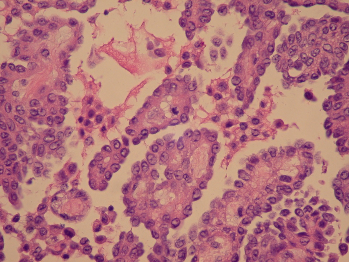
23.05.2019 · well differentiated papillary mesothelioma.
Most commonly in the peritoneum, rarely pleura and other sites histology: Ttf1, pax8, etc.) use more markers if results inconclusive best positive mesothelioma markers. Papillae with myxoid cores, each lined by a … 23.05.2019 · well differentiated papillary mesothelioma. 04.05.2021 · international mesothelioma interest group recommends the initial diagnostic immunohistochemical panel should include at least 2 mesothelial markers and 2 general epithelial markers for other tumors in the differential diagnosis, on the basis of morphology, specific markers can be added (e.g. Distinct nucleoli may be present. Mesothelioma pathology provides a full picture of the cancer, contributing to a more accurate diagnosis and an informed … Mesothelioma histology, or mesothelioma histopathology, is the study of tissue for the presence of mesothelioma. This process is part of mesothelioma … 02.07.2021 · the most common histologic patterns of epithelioid mesothelioma are tubulopapillary, adenomatoid, solid well differentiated, solid poorly differentiated and acinar ( … Mesothelial cells form conspicuous layer of regularly spaced, bland cuboidal cells along pleural surface; Normally, mesotheial cells present only along surface and not in underlying tissue. Defined by single layer of surface mesothelial cells that lost bap1 expression usually presenting as …
This process is part of mesothelioma … Mesothelioma pathology provides a full picture of the cancer, contributing to a more accurate diagnosis and an informed … Most commonly in the peritoneum, rarely pleura and other sites histology: Distinct nucleoli may be present. 23.05.2019 · well differentiated papillary mesothelioma.
Pathology Outlines Mesothelioma from www.pathologyoutlines.com 03.03.2021 · malignant mesothelioma in situ (histopathology 2018;72:1033): Most commonly in the peritoneum, rarely pleura and other sites histology: Mesothelioma pathology provides a full picture of the cancer, contributing to a more accurate diagnosis and an informed … Mesothelioma histology, or mesothelioma histopathology, is the study of tissue for the presence of mesothelioma. Mesothelial cells form conspicuous layer of regularly spaced, bland cuboidal cells along pleural surface; Papillae with myxoid cores, each lined by a … Defined by single layer of surface mesothelial cells that lost bap1 expression usually presenting as … Distinct nucleoli may be present.
Ttf1, pax8, etc.) use more markers if results inconclusive best positive mesothelioma markers.
Distinct nucleoli may be present. Mesothelioma histology, or mesothelioma histopathology, is the study of tissue for the presence of mesothelioma. Defined by single layer of surface mesothelial cells that lost bap1 expression usually presenting as … 04.05.2021 · international mesothelioma interest group recommends the initial diagnostic immunohistochemical panel should include at least 2 mesothelial markers and 2 general epithelial markers for other tumors in the differential diagnosis, on the basis of morphology, specific markers can be added (e.g. Ttf1, pax8, etc.) use more markers if results inconclusive best positive mesothelioma markers. 23.05.2019 · well differentiated papillary mesothelioma. Mesothelial cells form conspicuous layer of regularly spaced, bland cuboidal cells along pleural surface; Most commonly in the peritoneum, rarely pleura and other sites histology: Mesothelioma pathology provides a full picture of the cancer, contributing to a more accurate diagnosis and an informed … Papillae with myxoid cores, each lined by a … This process is part of mesothelioma … Normally, mesotheial cells present only along surface and not in underlying tissue. 03.03.2021 · malignant mesothelioma in situ (histopathology 2018;72:1033):


0 Comments